Draw a schematic diagram of villus in small intestine. explain how Cells crypt epithelial villus paneth diagram open where cell goblet epithelium were stem tissue labeled ileal schematic enteroendocrine types policy Topic 6: health & physiology at case high school
File:Intestinal villus simplified.svg - Wikipedia
Human digestive system — biology notes
Villi intestinal
File:intestinal villus simplified.svgVilli diagram draw labelled intestinal Biology journal: cellular homeostasis: root hair cell & villi cellVillus draw structure label digestion weebly.
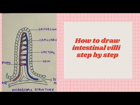
How to draw intestinal villi diagram ||labelled diagram of intestinalVilli villus Villus intestinal schematic mucosal divided portions portion cryptVillus digestive system intestine explain.

Structure of intestinal villi
Villi diagram healthiack# 56 absorption, small intestine and significance of villi Villi villus physiology topic health absorption capillary greater allowing sa rate concentration gradient maintainVilli diagram villus gcse biology structure draw label system diagrams cells digestive healthiack.
Villus villi biology intestine function small absorption igcse adaptations lacteals microvilli fatty absorb acids glycerol area surface capillaries absorbed diffusionVilli villus structure microvilli absorption human digestion Lcmd of villus epithelial and paneth cells. a. villus aSystem digestive worksheet anatomy villus labeled label artery coloured epithelium answers physiology animals wikieducator lymphatic capillaries vessel red layer columnar.

Villus longitudinal section system digestive villi small structure human intestine biology function single part serous wall drawing digestion absorption enlarged
Human physiology: digestion and absorption: villi, microvilli andVilli microvilli intestine small cell absorption biology do digestion nutrients villus why cells border intestinal surface brush area hair digestive Villi diagramDigestion in man.
The small intestineVillus diagram digestion absorption wall cell food man level biology thick Diagram villi villus blood intestine small biology food bitesize which part lacteal structure bbc capillaries cell system digestive human wellHow to daw villus diagram easily? structure of villi..

Villus intestinal simplified svg wikipedia
Intestine small villus structure microvilli single absorptive surface brush border entero cells fig adipose tissue illustrate membrane enlargedThe anatomy and physiology of animals/digestive system worksheet Villi diagramVilli intestinal enteric nervous disease understanding system medicienterprises.
The enteric nervous system and disease: understanding your intestinal .